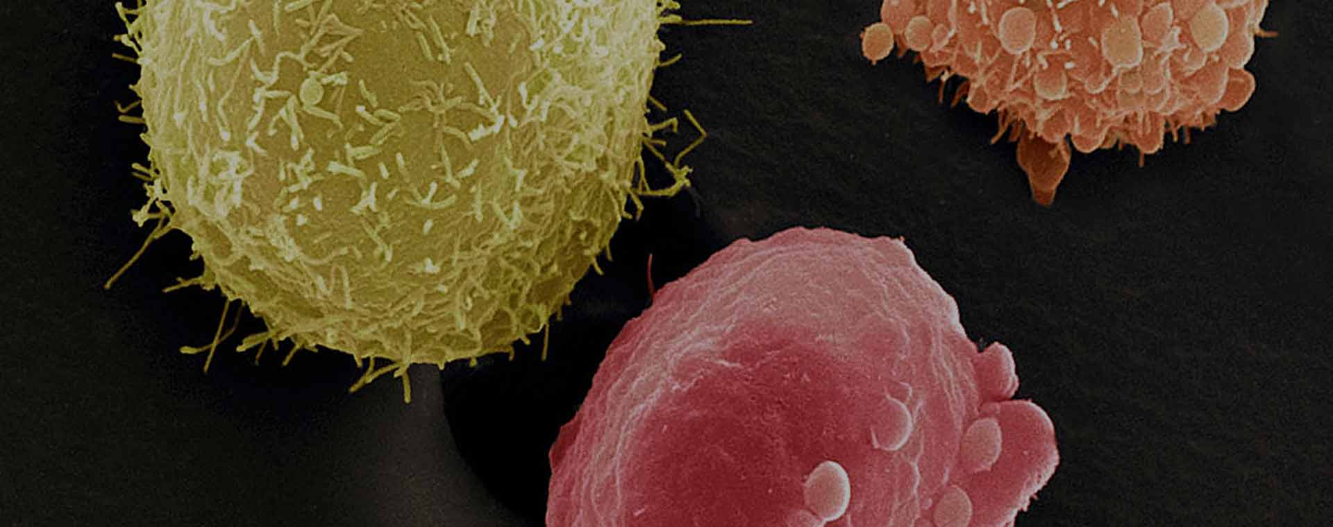
The majority of bladder cancers are low grade, non invasive or superficial tumours. These make up about 80% of tumours and although they have a high chance of recurrence they rarely progress to invasive or dangerous tumours. About 20% of tumours are invasive tumours which can be life threatening. Tumours can be high grade and non invasive and these tumours are also significantly more dangerous.

Bladder Cancer
The most common risk factor for bladder cancer is smoking.
The most common risk factor for bladder cancer is smoking. The risk increases with both daily number of cigarettes and the number of years one has smoked. There is also an increased risk from industrial exposure to aniline dyes and aromatic amines associated with textile and petrochemical industries.
Symptoms
By far the most common symptom is painless haematuria , blood in the urine.
Other symptoms involve changes to the normal urinary habit such as increased frequency or urgency or pain and burning. Recurrent infections can also be a sign of bladder cancer.
Diagnoses
Urine tests
A urine test will be sent for culture and microscopy and to detect blood in the urine.
A urine test may be performed to identify cancer cells called urine cytology. A negative test does not exclude cancer but a positive test usually indicates a high risk of high grade disease.
Imaging
Imaging- an Ultrasound of the bladder and kidneys will often be performed as well as a CT IVP which is a contrast study of the entire renal tract including the kidneys, ureters and bladder. This studies help in staging the tumour which gives an idea of the extent of the disease and whether or not it has spread out of the bladder.
Cystoscopy
A cystoscopy will be performed which is the passing of a telescope and camera into the bladder via the urethra to examine the lining. If a tumour or abnormality is seen in the bladder it will be biopsied or completely resected. The resected tumour will be sent for pathological examination. The results of the pathological examination will determine if it is a high grade or low grade tumour and will determine the depth of invasion of the tumour. These results will be the determining factor for treatment.
Treatment
The majority of tumours will be low grade non invasive tumours. I refer to these as “nuisance” tumours as they are not life threatening but need regular surveillance and follow up. These tumours will be treated with Cystoscopy and transurethral resection of the tumour followed by regular check cystoscopies to ensure they have not recurred.
At the time of first diagnosis a single dose of intravesical chemotherapy may be given which will decrease the rate of recurrence. This is a liquid that is placed in the bladder at the time of cystoscopy or via a urethral catheter within 24 hours of the cystoscopy.
If tumours are recurring often then a 6 week course of intravesical chemotherapy may be given between cystoscopies to decrease recurrence.
If tumours are non invasive but high grade they have a high risk of progressing to invasive tumours so as well as the regular cystoscopies they will require intravesical immunotherapy known as BCG. Like chemotherapy, this is a liquid that is placed in the bladder via a catheter and left in for an hour and then released. This is done weekly for 6 weeks and may require maintenance courses for 3 weeks every 6 months between cystoscopies for a period of 2 years. If these tumours recur whilst on BCG treatment or progress to invasive disease then removal of the bladder is considered.
In some cases of high grade or invasive disease that has not yet become muscle invasive removal of the bladder will be considered early as this gives a greater chance of cure.
If bladder cancer is muscle invasive it is a life threatening condition and aggressive treatment is necessary. The treatment options depend on the age and fitness of the patient and some tumour factors but generally the best chance of cure is complete removal of the bladder known as Radical Cystectomy. In males this also involves the removal of the prostate whilst in females it often involves removal of the uterus and part of the vagina.
After removing the bladder a urinary diversion is needed which is either in the form of an ileal conduit which is the most common and requires an external stoma or a neobladder, which is a longer and more complex procedure where a new bladder is made from small bowel and has no external drainage of urine leaving the body image intact.
Systemic chemotherapy may be considered before or after a cystectomy depending on the extent of the disease and an alternative to surgery is a combination of Chemotherapy and Radiotherapy.
Cystectomy is very major surgery and the procedure and side effects of the procedure will be discussed with you in detail if you require this treatment. In both males and females sexual function will be affected.
All of these cases will be discussed in a Multi Disciplinary team meeting involving urologists, medical and radiation oncologists. For those cases where the disease has already spread outside the bladder known as metastatic disease, it is no longer potentially curable and palliative therapy will involve the medical and radiation oncologists.


